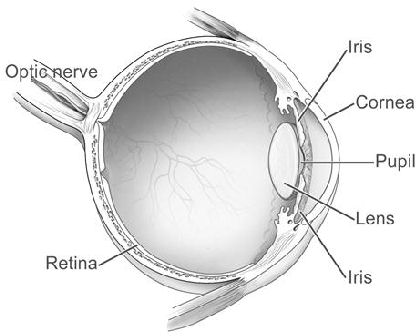
Glossary / Anatomy of the Eye
- Cornea
- The cornea is the eye's outermost layer. It is the clear, dome-shaped surface that covers the front of the eye.
- AMD-Dry Form
- The dry form of age-related macular degeneration (AMD) occurs when the light-sensitive cells in the macula slowly break down, gradually blurring central vision in the affected eye. Over time, as less of the macula functions, central vision is gradually lost in the affected eye.
- AMD-Wet Form
- The wet form of age-related macular degeneration occurs when abnormal blood vessels behind the retina start to grow under the macula. These new blood vessels tend to be very fragile and often leak blood and fluid. The blood and fluid raise the macula from its normal place at the back of the eye. Damage to the macula occurs rapidly.
- Floaters
- Floaters are tiny spots, specks, flecks and "cobwebs" that drift aimlessly around in your field of vision.
- Lens
- The lens is the clear part of the eye that helps to focus light, or an image, on the retina.
- Macula
- The macula is the part of the eye that allows you to see fine detail. The macula is located in the center of the retina.
- Optic Nerve
- The optic nerve sends visual information from the retina to the brain.
- Retina
- The retina is the light-sensitive tissue at the back of the eye. The retina instantly converts light, or an image, into electrical impulses. The retina then sends these impulses, or nerve signals, to the brain.

Courtesy of the National Eye Institute, National Institutes of Health